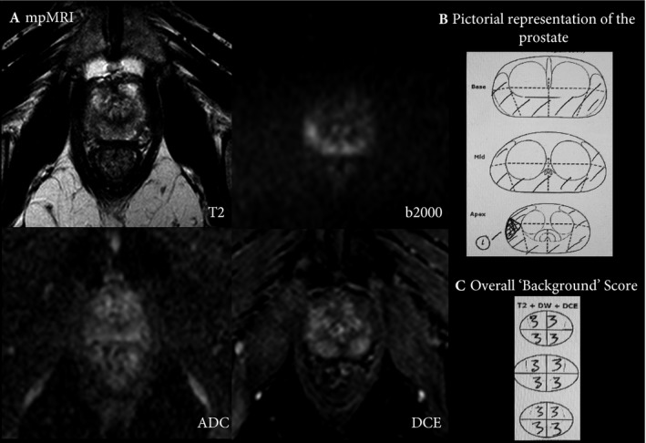
Figure 1.

(A) Shows the mpMRI of a 62‐year‐old man, with a PSA level of 4.4 ng/mL and a gland volume of 25 mL at the level of the mid‐gland to apex region. On T2W imaging, there is diffuse and patchy low T2 signal and a lower T2 signal at the right lateral gland, with an equivocal high signal focus on diffusion high b value at 9 o'clock and corresponding equivocal low ADC signal with bilateral enhancement on DCE. The focal lesion (represented by number 1 in 1. (B) was reported with a Likert‐assessment of 3/5. Besides, the remainder of the gland was also assessed with the whole prostate divided into quarters for Likert assessment (C). Each quarter was reported as a ‘Likert‐assessment’ 3/5. The background changes scored 3 are represented by the shaded area in B. Upon transperineal template mapping biopsy, the prostate was found to harbour adenocarcinoma Gleason 3+4, (40% biopsy core involvement) at the right posterior apex, focal high‐grade prostatic intraepithelial neoplasia at the left posterior apex and Gleason 3+3, at eight different sites within the prostate (10–40% biopsy core involvement).
