Fig. 8.
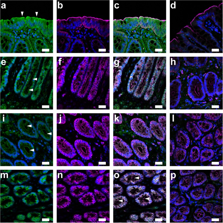
Immunolocalization of α- and β-subunits of KCNQ channels in the rat rectal colon. a Fluorescence of KCNQ2 on the luminal membranes of surface cells (arrowheads). b Fluorescence image of ezrin. c Overlay image of a and b. d Overlay image of ezrin and green fluorescence with KCNQ2 antibody pre-absorbed with the control peptide antigen. e Fluorescence of KCNQ4 on the luminal membrane of crypt cells (arrowheads). Fluorescence images of ezrin (f), the overlay (g), and negative control with the pre-absorbed KCNQ4 antibody (h). i Fluorescence of KCNE3 on the luminal membrane of crypt cells (arrowheads). Fluorescence images of ezrin (j), the overlay (k), and negative control with the pre-absorbed KCNE3 antibody (l). Fluorescence of KCNQ1 (m), ezrin (n), and the overlay (o) in crypts. Arrowheads show the luminal membrane of crypt cells. p Negative control with the pre-absorbed KCNQ1 antibody. DAPI was used to stain nuclei (blue). Bars = 20 μm

