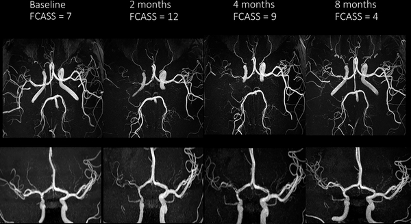
Figure 2.
Cerebral MRA images demonstrating evolution of a typical case of FCAi: axial views (upper row) and frontal views (lower row) at four time points. Baseline images demonstrate mild irregularity of the right supraclinoid ICA and M1 segment of the middle cerebral artery. The 2-month images show progression to severe stenosis; the 4- and 8-month images demonstrate subsequent improvement.

