Fig. 6.
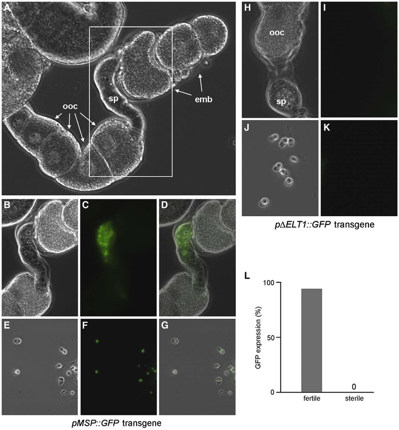
Expression of an ELT-1 target transgene in sperm. (A) DIC image of dissected gonad from pMSP::GFP transgenic hermaphrodite. (B–D) Higher magnification of boxed region in panel A showing DIC (B), GFP (C), and composite (D) images. (E–G) Spermatozoa from dissected spermatheca of pMSP::GFP transgenic hermaphrodite by DIC (E), GFP (F), and composite (G). (H–K) Dissection of pΔELT1::GFP transgenic hermaphrodites showing gonad (H, I) or spermatozoa (J, K) by DIC or GFP fluorescence, respectively. (L) Loss of GFP expression in sterile hermaphrodites. 1440 homozygous elt-1(ok1002); pMSP::GFP hermaphrodites containing the elt-1+rol-6(su1006) extrachromosomal array were individually screened for sterility then visualized for GFP in sperm (fertile, N=200; sterile, N=8). ooc, oocytes in the proximal arm of the uterus; sp, spermatheca; emb, embryos in the uterus.

