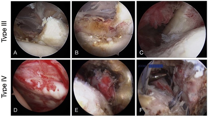
Figure 8.
Arthroscopic fixation of type III and type IV tears. (A-C) Type III tear. Note the disruption of the superior tendinous portion in (A) and continuity of the muscle portion in (B). (C) Fixation of type III tear. (D-F) Type IV tear. (D) Before mobilization of tendon. (E) After mobilization of tendon; note how far back the tendon is retracted. (F) Suture fixation of the tendon with multiple anchors.

