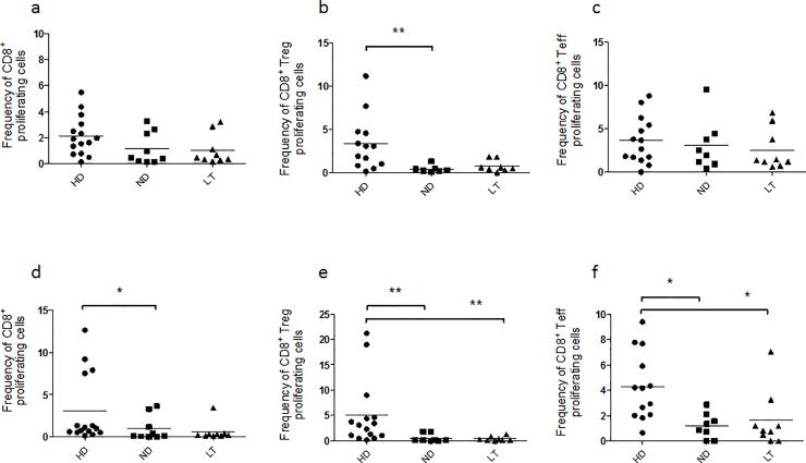
Fig 5. Proliferative responses of cell subsets under study in HD, ND and LT T1D patients after three days of PMA/ionomycin stimulation.
CMFDA-labeled PBMC from healthy controls and T1D patients were stimulated with PMA/ ionomycin for three and five days and subsequently stained for flow-cytometry analysis. Graphs show the frequency of CD8+, CD8+ Treg, CD8+ Teff proliferating cells after 3 (a-c) and 5 days (d-f) of PMA/ionomycin stimulation. Proliferation was evaluated as percentage of CMFDA-low cells relative to the subset analyzed after stimulation over the percentage of CMFDA-low cells of the same subset in RPMI unstimulated cultures. For the investigation present in figure, 14 HD, 9 ND and 9 LT samples were studied.

