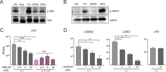
Fig 3. Differential utilization of upstream autophagy components by RNA viruses.
(A and B) Cells were treated with rapamycin or infected with PV (6 hpi), DENV (24 hpi), or ZIKV (24 hpi). Cell lysates were harvested and immunoblotted with phospho-S6K or phospho-AMPK antibodies. (C) Cells were treated with 0.5, 5, or 50 μM of MBL56 and infected with PV at an MOI of 0.1 PFU/cell for 6 hours. (D) WT cells were pretreated with 1, 5, or 10 μM of SAR405 and infected with DENV or ZIKV at an MOI of 0.1 PFU/cell for 24 hours (DENV) or 48 hours (ZIKV). WT cells were pretreated with 10 μM of SAR405 and infected with PV at an MOI of 0.1 PFU/cell for 6 hours. All data are represented as mean +/− SEM. *Indicates significant P value of <0.05, **P value < 0.01, ***P value < 0.001, ****P value > 0.0001 by an unpaired t test. See also S3 Data. AMPK, AMP-activated protein kinase; DENV, dengue virus; hpi, hours post infection; MOI, multiplicity of infection; PFU, plaque-forming units; PV, poliovirus; WT, wild-type; ZIKV, Zika virus.

