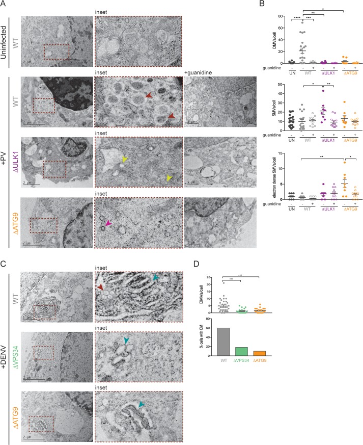
Fig 4. Virally induced membrane rearrangements are altered in cells that lack individual autophagy components.
(A and B) HeLa cells were infected with PV or DENV at an MOI of 10 PFU/cell. Indicated cells were treated with 2 mM guanidine during the infection period. Cells were fixed at 6 hpi (PV) or 24 hpi (DENV) and subjected to high-pressure freezing and freeze substitution. Images were collected on a TEM microscope. Representative images are shown. Red arrowheads indicate DMVs, yellow arrowheads indicate SMVs, pink arrowheads indicate electron-dense SMVs, blue arrowheads indicate CM. Quantification of cellular structures was done on blinded images, and >10 cells per condition were counted. All data are represented as mean +/− SEM. *Indicates significant P value of <0.05, **P value < 0.01, ***P value < 0.001, ****P value > 0.0001 by a Mann–Whitney test. See also S4 Data. CM, convoluted membranes; DENV, dengue virus; DMV, double-membraned vesicles; HeLa, human epithelial-derived cell line; hpi, hours post infection; MOI, multiplicity of infection; PFU, plaque-forming units; PV, poliovirus; SMV, single-membraned vesicles.

