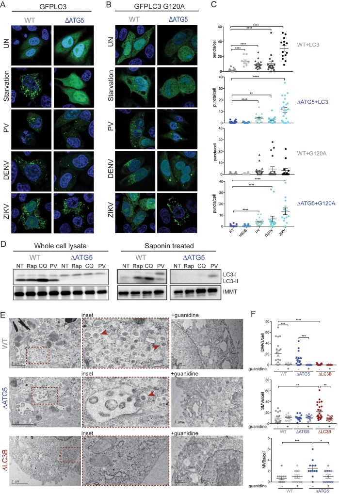
Fig 5. LC3 is recruited to membranes independent of lipidation during viral infection.
(A and B) HeLa cells were transfected with GFP–LC3 or GFP–LC3–G120A for 48 hours. Cells were either starved for 2 hours or infected at an MOI of 10 PFU/cell with PV (6 hours), DENV or ZIKV (24 hours) and fixed for visualization by confocal microscopy. (C) Puncta per cell were counted for each condition; n = >10 cells. (D) HeLa cells were treated with Rap, CQ, or infected with PV at an MOI of 10 PFU/ml for 6 hours. Lysates were harvested with or without saponin and run on an SDS PAGE gel. Immunoblots were stained for LC3 and the membrane-associated IMMT. (E) HeLa cells were infected with PV at an MOI of 10 PFU/cell and subjected to high-pressure freezing and freeze substitution for visualization on a TEM microscope. Representative images are shown. Red arrowheads indicated DMVs. (F) Cell structures were quantified on blinded images; n = >15 cells. All data are represented as mean +/− SEM. *Indicates significant P value of <0.05, **P value < 0.01, ***P value < 0.001, ****P value > 0.0001 by a Mann–Whitney test. See also S5 Data. CQ, chloroquine; DENV, dengue virus; DMV, double-membraned vesicle; GFP, green fluorescent protein; HeLa, human epithelial-derived cell line; IMMT, inner membrane mitochondrial protein; LC3, light-chain 3; MOI, multiplicity of infection; PFU, plaque-forming units; PV, poliovirus; Rap, rapamycin.

