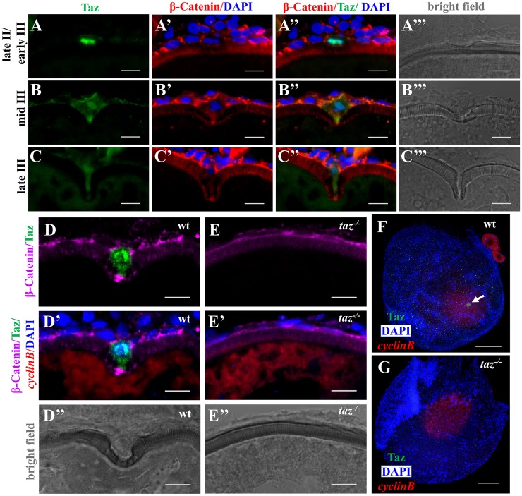
Fig 5. Taz is highly enriched in micropylar cells.
(A-C’”) Immunofluorescence of Taz and β-Catenin in sectioned wild type oocytes at various stages: late II/ early III (A-A’”, n = 14), mid III (B-B’”, n = 14) and late III (C-C’”, n = 13). β-Catenin and DAPI label the cell membrane and nucleus, respectively. While basal levels are detected in follicle cells, in the micropylar cell high levels of Taz are detected, predominantly in the nucleus (A-C, A”-C”). Taz expression levels decrease as the micropylar cell develops (A-C, A”-C”). The micropylar cell undergoes dramatic shape change from flattened to ‘nail’-like to perforate the developing vitelline envelope (A’-C’, A”-C”). Bright field images show the gradual invagination of the vitelline envelope by the protruding micropylar cell (A’”-C’”). (D-E”) Immunofluorescence of Taz and β-Catenin in sectioned wild type and taz-/- oocytes at stage III after in situ hybridization to detect cyclinB. In wild type ovaries, the high Taz expressing micropylar cell sits on the top of the oocyte animal pole, marked by cyclinB, and perforates the vitelline envelope (D-D”, n = 9). No Taz is detectable in taz-/- oocyte (E), and neither the micropylar cell around the animal pole nor an invagination on the vitelline envelope is observed in mutant oocytes (E-E”, n = 11). (F-G) Immunofluorescence to detect Taz and in situ hybridization for cyclinB in wild type and taz-/- oocytes at stage III. A single high Taz expressing micropylar cell is located at the animal pole of wild type oocyte (F, n = 13), but is not found in taz-/- ovaries (G, n = 17). Two stage I oocytes, ubiquitously expressing cyclinB, are found adjacent to the stage III oocyte in figure F. White arrow, the micropylar cell indicated by high Taz expression. Scale bar, 100 μm in F and G and 10 μm in others.

