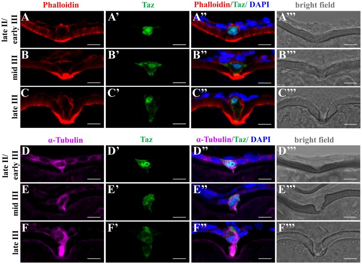
Fig 7. Taz may regulate cytoskeletal dynamics in the micropylar cell.
(A-F’”) Immunofluorescence shows Taz and F-actin (A-C’”, n = 32) or α-Tubulin (D-F’”, n = 19) in sectioned ovaries; besides the invagination on the vitelline envelope, the expression of Taz and the shape of the micropylar cell changes with the growth of the micropylar cell; Phalloidin labelled F-actin bundles are found gradually deposited in the leading tip in the cytoplasmic extension of the micropylar cell and the part of oocyte cortex contacting the micropylar cell (A-C’”), and α-Tubulin is enriched in the cytoplasmic extension of the micropylar cell (D-F’”). Scale bar, 10 μm.

