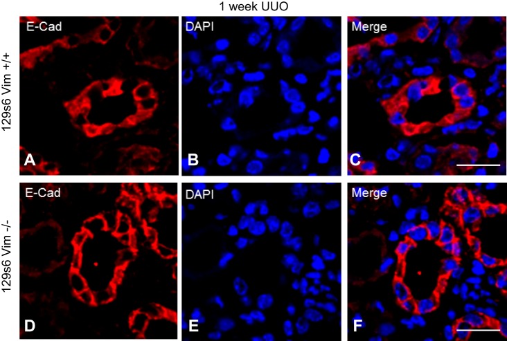
Fig. 5.
Vimentin (vim) −/− mice undergoing unilateral ureteral obstruction (UUO) display no difference in E-cadherin in proximal renal tubules following UUO. OCT-embedded kidneys following UUO underwent immunofluorescence with antibodies directed against E-cadherin and DAPI. All images were obtained on with a Leica DMI4000 B confocal microscope and analyzed using Leica advanced fluorescence application suite. E-cadherin localization 1 wk following UUO in vim −/− mice reveals a strong peripheral staining pattern along the cell membrane (D and F), whereas wild-type (WT) mice E-cadherin is seen both in at the cell membrane as well as the cytoplasm (A and C). Size bars = 10 μm.

