Fig. 6.
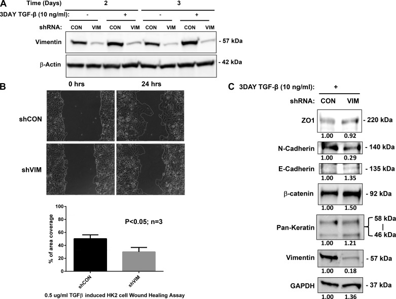
Vimentin (vim) mediates transforming growth factor (TGF)-β-induced epithelial to mesenchymal transition (EMT) of human renal tubular epithelial cells (A). Sequence of shRNA to human vim. shRNA specially targeting (shVIM) downregulated vim expression efficiently in human proximal renal tubular (HK-2) cells (B). Two and 3 days following induction with TGF-β, control HK-2 cell lines express vim, whereas shVIM cell lines (VIM) do not express vim in appreciable amounts. All lanes were normalized to β-actin to ensure proper loading. Time lapse phase contrast microscopy was used to visualize wound healing in control cells (shCON) and shVIM cells following TGF-β induction (C). Quantification of cellular migration was carried out by measuring the changes in the wound area after 24 h (D). There was a ~30% reduction in migration following EMT induction in shVIM cell lines. Experiments were performed in triplicate and P values calculated. Immunoblotting using antibodies against vim, pan-keratin, and EMT markers, including zona occludens (ZO)-1, N-cadherin, E-cadherin, and β-catenin was carried out on shVIM HK-2 cells following TGF-β induction (E). Signals were normalized to GAPDH to account for variability in loading and then expressed as fold changes of the negative control, shCON. shVIM HK-2 cells demonstrate “sheet-like” motility following TGF-β stimulation. Vim-silenced HK-2 cells were treated with TGF-β and then underwent wound healing assay. Time lapse phase contrast microscopy for 24 h demonstrate sheet-like movement when compared with control cell lines. Time lapse assembled from 1-h images over 24-h period. Supplemental Material for this article is available online at the Journal website.

