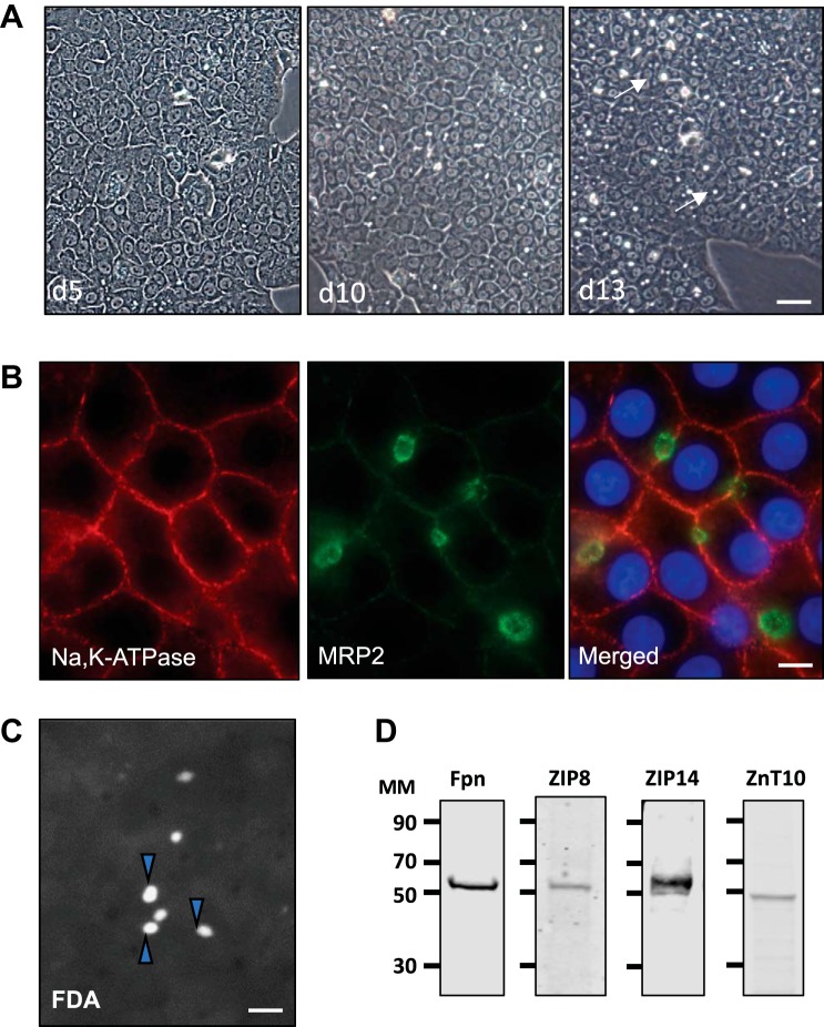
Fig. 1.
WIF-B cell polarization and manganese (Mn) transporter levels. A: WIF-B cells were seeded at a density of 1 × 10⁶ cells on 10-cm2 dishes, and cultures were observed by phase contrast microscopy after the indicated days in culture. Bile canalicular compartments (BCs) (phase bright structures) are indicated by arrows (×10 objective; bar = 40 µm). Representative micrographs of culture conditions observed throughout the time frame of all experiments presented in the paper are shown. B: indirect immunofluorescence (IF) microscopy of WIF-B cells after 13 days in culture shows immunoreactivity for Na,K-ATPase on the basolateral membrane and apical staining for the bile canalicular marker MRP2; nuclei indicated by DAPI (×63 oil objective; bar = 10 µm). IF studies were carried out with cells examined from duplicate wells, and each experiment was performed on at least three separate occasions with the same observations. C: functional integrity of WIF-B cell BCs after 12 days in culture was determined upon incubation with 0.1 µg/ml fluorescein diacetate (FDA) for 20 min at 37°C. After washing, live cell images were captured within 10 min (×20 objective; bar = 20 µm). FDA experiments were carried out with triplicate wells and on three separate occasions with similar results. Arrowheads indicate BCs that have accumulated fluorescein. D: Western blot analysis of cell lysates prepared from WIF-B cells grown for 14 days in culture. Immunoreactivity was detected for ferroportin (Fpn), ZIP14, ZIP8, and ZnT10. d5, day 5; d10; day 10; d13, day 13; MM, molecular mass (kDa).

