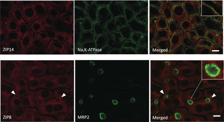
Fig. 6.
ZIP14 and ZIP8 distribution in WIF-B cells. WIF-B cells were grown on coverslips for 14 days for maximal bile canalicular compartment (BC) density and indirect double immunofluorescence (IF) performed to image Na,K-ATPase and ZIP14 or ZIP8 and MRP2. ZIP14 colocalized with Na,K-ATPase (inset; Pearson’s = 0.456). ZIP8 staining did not associate with MRP2 structures (arrows; inset, Pearson’s = 0.078). IF results were obtained on three separate occasions with duplicate wells prepared on each day.

