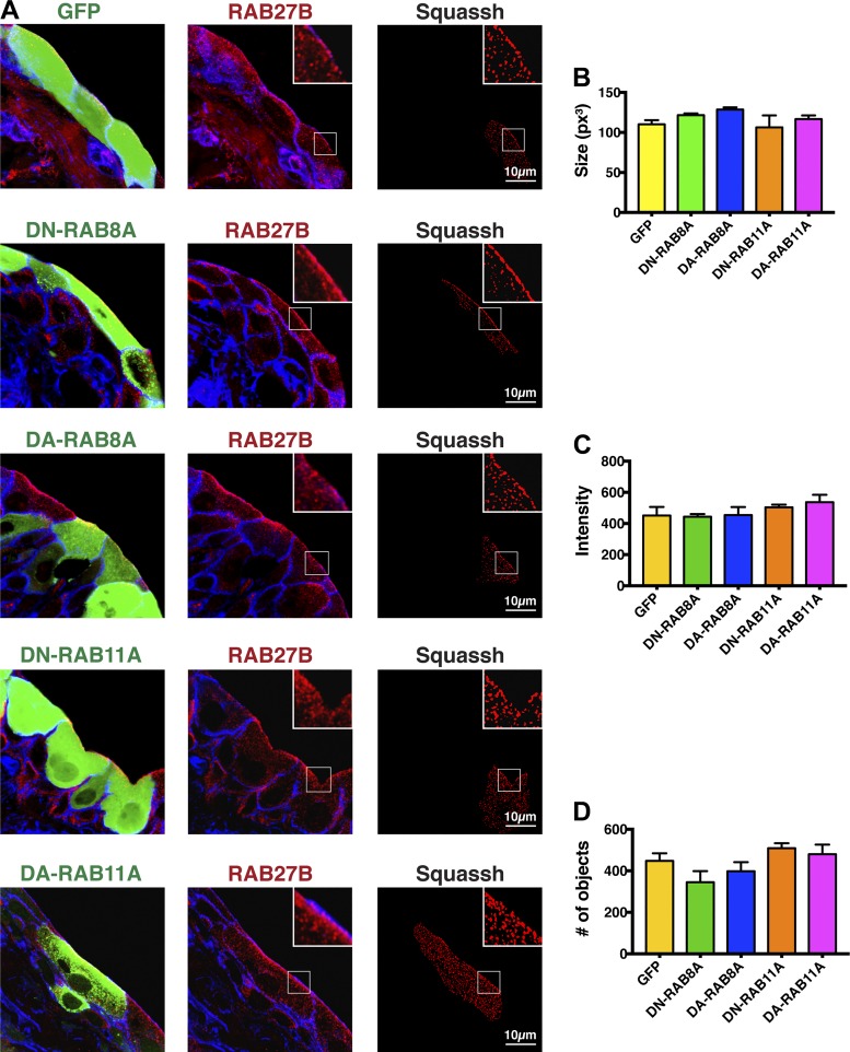
Fig. 6.
Effect of expressing mutants of RAB8A and RAB11A on the distribution, size, intensity, and number of RAB27B vesicles. Bladders were transduced with adenoviruses encoding GFP alone (control), or GFP-tagged dominant-negative (DN) RAB8A, or RFP-tagged dominant-active (DA) RAB8A, GFP-tagged DN-RAB11A, or GFP-tagged DA-RAB11A. A: confocal analysis. The column at left shows expression of the indicated protein (green), the column at middle shows the distribution of RAB27B in the tissue (red), and the column at right shows the Z stack after cell masking and Squassh segmentation analysis of the RAB27B channel. Summary statistics for the size (B), intensity (C), and number (D) of RAB27B vesicles in cells expressing the indicated protein. None of the values are significantly different from GFP controls or from one another (assessed using ANOVA).

