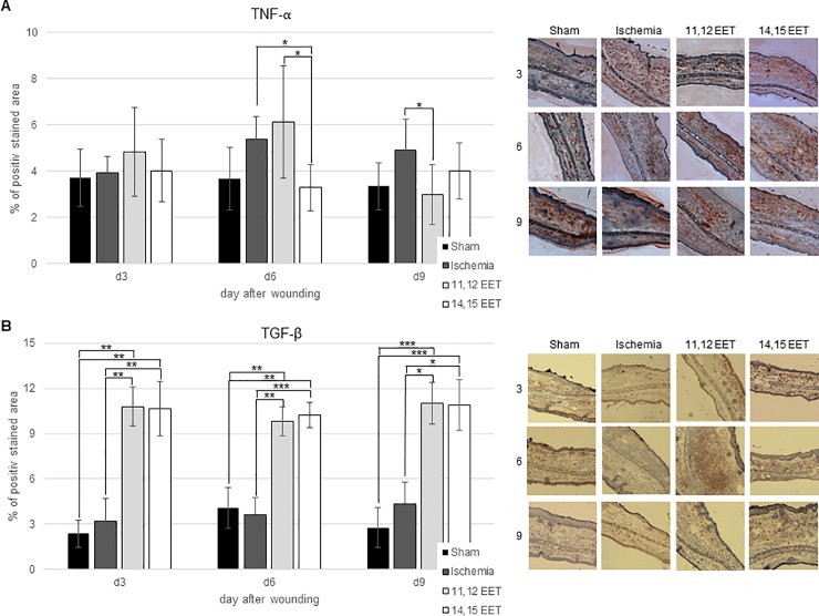
Fig 4.
A Percentage of TNF-α positive area on day 3, 6, and 9 after wounding of control, ischemic and 11,12 as well as 14,15 EET treated ischemic wounds. On the right representative pictures of immunohistological staining. B Percentage of TGF-β positive area on day 3, 6 and 9 after wounding of control, ischemic and 11,12 as well as 14,15 EET treated ischemic wounds. On the right representative pictures of immunohistological staining (data is shown as mean ± SD; n = 8). *p<0.05, ***p<0.001.

