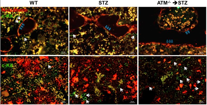
Figure 1: Localization of c-Kit+ cells in mouse bone marrow.
Upper panel: immunofluorescence staining of N-cadherin (red) and c-Kit (green) in demineralized mouse femurs. Some c-Kit+ cells (white arrow) localized to endosteal niche (blue arrow) are defined as long-term repopulating (LTR)-hematopoietic stem cells (HSCs); Lower panel: mouse femurs stained for VE-cadherin (red) and c-Kit (green). c-Kit+ cells (white arrow) located at vascular niche are defined as short-term repopulating (STR)-HSCs. Representative images showing a reduced number of LTR-HSCs and increased STR-HSCs/LTR-HSCs in the STZ-induced diabetic bone marrow. ATM−/− intensified diabetes-mediated defects of LTR- and STR-HSCs imbalance in the bone marrow. Abbreviations: WT, wild type; STZ, streptozotocin; ATM, ataxia telangiectasia mutated.

