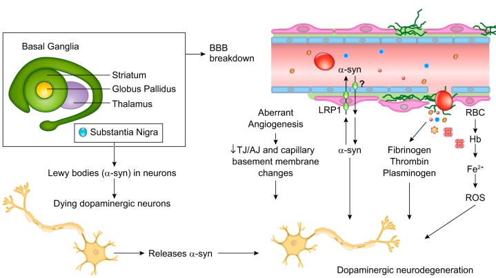
FIGURE 8.
Blood-brain barrier breakdown and dysfunction in sporadic Parkinson’s disease. Vascular dysfunction occurs throughout the basal ganglia of Parkinson’s disease (PD) patients, consisting of blood-brain barrier (BBB) breakdown and dysfunction, as shown by dynamic contrast-enhanced magnetic resonance imaging (DCE-MRI), T2*-MRI and susceptibility-weighted imaging (SWI)-MRI demonstrating microbleeds, and diminished active efflux of xenobiotics and other potential toxins, as indicated by verapamil-PET. Aberrant angiogenesis with increased number of endothelial cells, decreased tight junction (TJ) and adherens junction (AJ) proteins, and capillary basement membrane changes have been shown both in humans with PD and animal models. BBB breakdown can lead to neurotoxic accumulates of fibrin(ogen), thrombin, and plasmin(ogen), and red blood cell (RBC) extravasation, release of hemoglobin (Hb) and iron (Fe2+) causing reactive oxygen species (ROS), which all can injure dopaminergic neurons. Concurrently, localized to the substantia nigra pars compacta, Lewy bodies form from filamentous and oligomeric α-synuclein (α-syn) that accumulate within dopaminergic neurons. Recent studies suggested that α-syn can cross the BBB and contribute to α-syn pool in the brain, and is also cleared from brain across the BBB via LRP1-mediated transcytosis.

