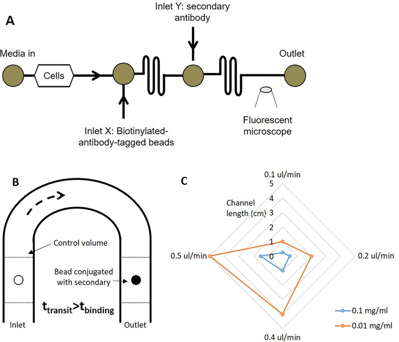
Figure 1.

(A) Schematic of the device to carry out the continuous-flow ELISA. Cell perfusate is carried the first mixing channel where it conjugates with the biotinylated beads. The antigen-tagged beads and the secondary antibody conjugate in the second mixing channel. The fluorescently labeled beads are detected under a fluorescence microscope at the outlet, (B) Schematic of the analytical model, and (C) Plot representing the relationship between the channel dimensions, flow rate, and the concentration of the antibody.
