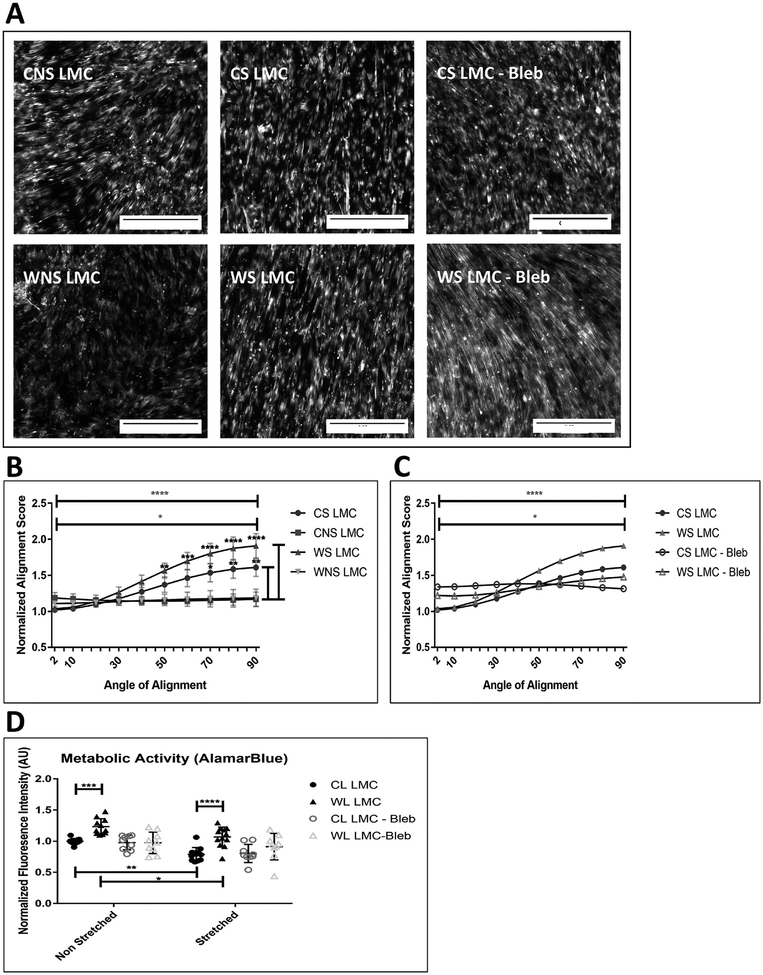
Figure 5: Impact of cyclic stretching on the alignment and metabolic activity in CL and WL LMCs.
(A) Representative images of F-actin fiber orientation of stretched (CS) and non-stretched (CNS) CL LMCs, and stretched (WS) and non-stretched (WNS) WL LMCs taken after 1 week. Scale bar is 0.25mm. (B) Quantification of F-actin fiber alignment for stretched and non-stretched LMCs. Preferential orientation perpendicular to strain (along 90o axis) is shown by comparing alignment score at 2o to alignment score at 90o for WS LMCs (top bar) and CS LMCs (2nd bar). Comparisons along the side are between the CS vs CNS LMCs and the WNS vs WS LMCs respectively. Error bars reflect standard error. There was no significant difference in the response between control and wounded LMCs (C) Blebbistatin (5μM) inhibited preferential alignment along 90o axis after cyclic strain. Error bars are removed for clarity. (D) Impact of cyclic stretching on metabolic activity of LMCs cultured in Standard DMEM, and DMEM supplemented with Blebbistatin (5μM). *: p<0.05, **: p<0.01, ***: p<0.001, ****: p<0.0001

