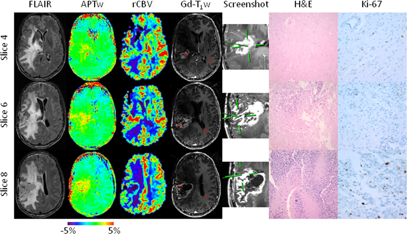
Fig. 2.
Anatomical, CBV, and APTw MR images, biopsied sites, and histology images for a patient with a treated GBM in the right parietal lobe (Case 2). Fifteen APTw slices were acquired. Three specimens were obtained from three different sites within different slices with distinct APTw signals, all with strong Gd enhancement. The specimen from Slice 4 was targeted at an APTw-isointense area (green; 1.62%), with an rCBV of 1.11, compared with CNAWM. The H&E-stained slice shows necrosis and a thickened vessel wall caused by hyalinization (Cellcount 677 cells/FOV and proliferation 2.7%). The specimen from Slice 6 is from a mildly APTw-hyperintense spot (yellow; 1.81%), with an rCBV of 2.12. The specimen shows predominant necrosis with limited residual tumor cells (Cellcount 876 cells/FOV and proliferation 5.1%). The specimen from Slice 8 was obtained from an APTw-hyperintense spot (yellow to red; 3.29%), with a high rCBV of 1.87. The specimen has obvious anaplasia, multinucleated tumor cells, and pseudopalisading necrosis (Cellcount 1673 cells/FOV and proliferation 11.2%). The use of APTw image-directed biopsy can potentially reduce the randomness of surgical decisions due to tumor heterogeneity.

