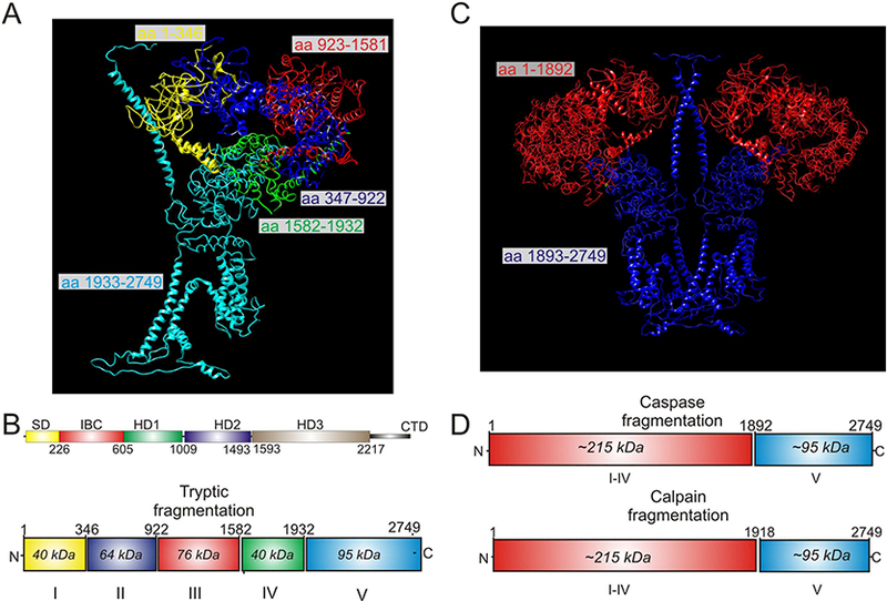
Figure 2.

Proteolytic fragments mapped to the Cryo-EM structure of R1. A and C are based on pdb 3JAV. A depicts a single subunit of R1 in which the five tryptic fragments generated by limited trypsin digestion are color coded. B shows these fragments in the linear structure. C Depicts a dimer of R1 in which the soluble (red) and membrane fragments (blue) formed by calpain and caspase activity in vivo are shown. D shows the calpain and caspase fragments in the linear structure of R1.
