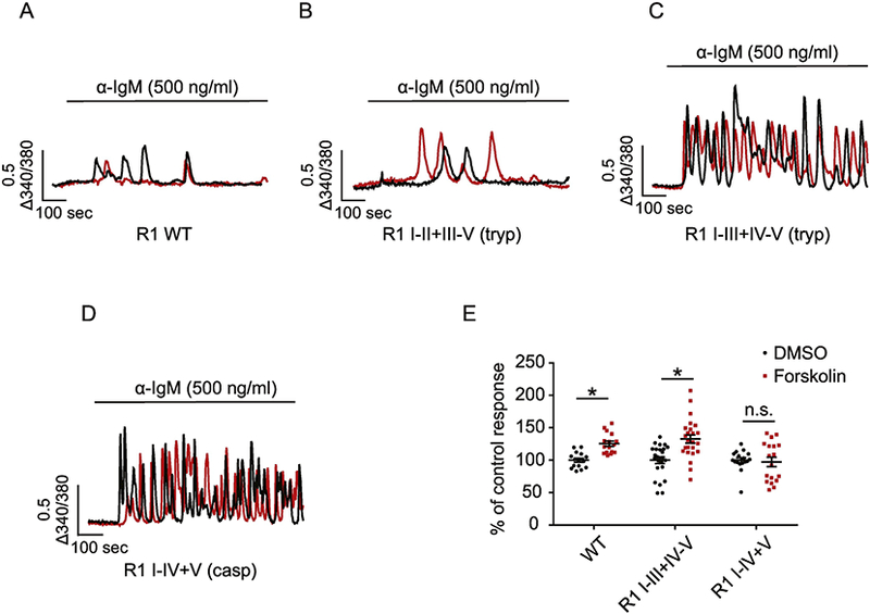
Figure 3.

Region-specific proteolysis regulates R1 activity. Single cell Ca2+ imaging was performed using cells expressing DT$) 3KO cells stably expressing R1 WT (A), R1 I-II+III-V (tryptic fragments) (B), R1 I-III+IV-V (tryptic fragments) (C) and R1 I-IV+V (caspase fragments) (D). Cells were loaded with fura-2AM followed by anti-IgM stimulation, which cross-linked the cell surface B cell receptors and induced continuous production of intracellular IP3. In response to anti-IgM stimulation, cells expressing R1 WT (A) and R1 I-II+III-V (tryptic fragments) (B) evoked few Ca2+ transients while cells expressing R1 I-III+IV-V (tryptic fragments) (C) and R1 I-V+V (caspase fragments) (D) exhibited robust Ca2+ oscillations. Two representative single cell calcium traces (black and red) were shown for each condition. In (E), pre-incubation of cells with 20 μM forskolin for 2 min activated PKA and phosphorylated R1. This resulted in significant increases in the amplitude of Ca2+ signals mediated by the full length R1 and fragmented R1 I-III+IV-V, but not R1 I-IV+V. Each point represents one experiment. * indicates p< 0.05. Adapted from [15] with permission.
