Fig. 3.
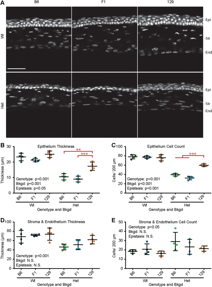
Epistasis influenced corneal epithelial thickness and cell counts. a Confocal scans of Pax6+/+ (Wt) and Pax6Sey/+ (Het) corneas on three genetic backgrounds (bkgds), C57BL/6J (B6), B6129F1 (F1), and 129S1/SvImJ (129) were examined. Quantification of (b) cornea epithelium (Epi) thickness, (c) Epi cell count, (d) stroma (Str) plus endothelial (End) thickness, and (e) Str plus End cell count, revealed that Pax6 genotype influenced all measurements. Additionally, epistasis between Pax6 genotype and bkgd influenced Epi thickness and cell count. Post hoc analysis of epistasis, indicated on graphs (red), revealed that Het 129 corneas had significantly thicker Epi than Het B6 and Het F1 corneas (**p < 0.005, and ***p < 0.001, respectively), and that Het 129 corneas had more Epi cells than Het B6 and Het F1 corneas (p < 0.001 for both). Gray staining (Hoechst), not significant (N.S.), scale bar = 50 µm. Statistical significance was determined using ANOVA, post hoc analysis was performed using Fisher’s LSD with Bonferroni’s correction for multiple comparisons. Each dot represents an individual eye and error bars represent the mean ± the standard deviation

