Figure 1.
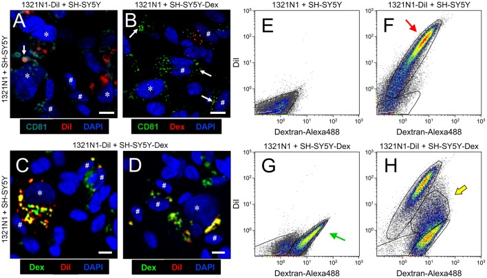
Astrocytes and neurons exchange CD81-positive material. (A–D) Single plane confocal microscopy images extracted from Z-stacks obtained from co-cultures of: 1321N1 astrocytes labeled with DiI and unlabeled SHSY5Y neurons (A); unlabeled 1321N1 and SH-SY5Y labeled with Dextran-Alexa 488 (B); 1321N1 labeled with DiI and SH-SY5Y labeled with Dextran-Alexa 488. (C,D) Immunocytochemistry with the extracellular vesicle (EV) marker CD81 is shown in (A,B). Arrows point to colocalizing signals. Asterisks and pound signs mark identified nuclei from astrocytes and neurons respectively. N = 20 confocal stacks/condition. Calibration bars: 10 μm. (E–H) Flow cytometry analyses plotting the DiI and Dextran-Alexa 488 signals in the different co-culture conditions. The red arrow in (F) indicates the DiI-1321N1 population, and the green arrow in (G) points to the Dextran-Alexa 488-SHSY5Y population. A double labeled cell population appears when both astrocytic and neuronal cells are labeled (H, marked with a yellow arrow).

