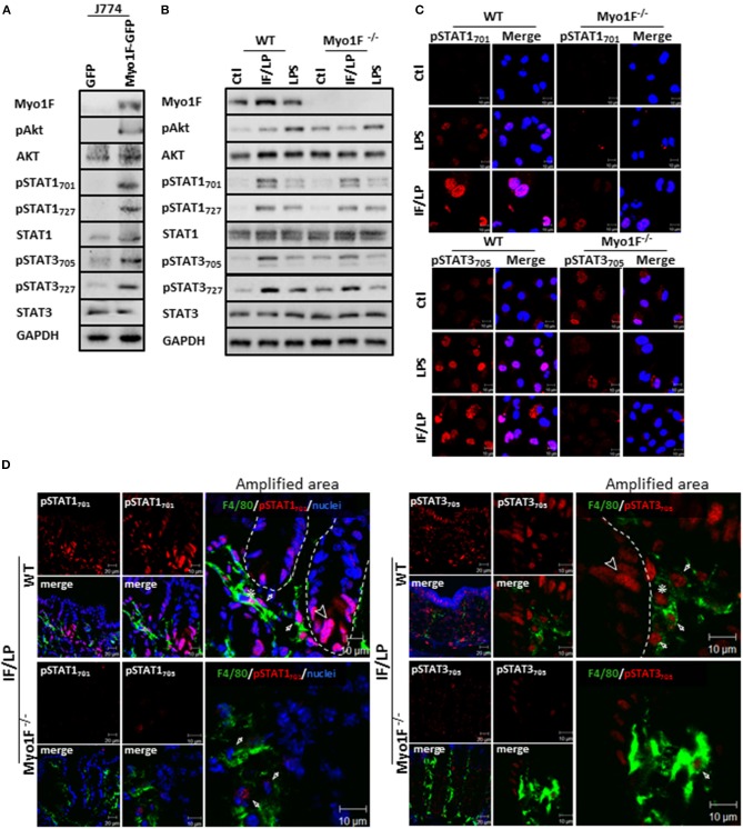
Figure 3.
Myo1F mediates activation and cellular localization of Akt/STAT signaling in macrophages. (A) Myo1F, pAkt473, AKT, pSTAT701, pSTAT727, STAT1, pSTAT3705, pSTAT3727, and STAT3 were analyzed in cell lysates of J774 cells expressing Myo1F-GPF or GFP. Cells were platted at confluence for 12 h before lysis. GAPDH was used as loading control. n = 3. (B) Myo1F, pAkt473, Akt, pSTAT701, pSTAT727, STAT1, pSTAT3705, pSTAT3727, and STAT3 were studied in WT or Myo1F−/− BMM. Cell lysates were carried out in macrophages control or stimulated for 5 h with IFN-γ (20 ng/ml)/plus LPS (1 μg/ml) stimulation or LPS alone (1 μg/ml). GAPDH was used as loading control. n = 3. (C) Immunofluorescence staining for pSTAT1 and pSTAT3 (red) was performed in WT or Myo1F−/− BMM platted in glass coverslips. Cell were fixed after 5 h stimulation with IFN-γ (20 ng/ml)/plus LPS (1 μg/ml), LPS (1 μg/ml) or carrier alone (Ctl). Nuclei were stained with Dapi (blue). Scale bar 10 μm. (D) Representative image of immunofluorescence staining for F4/80 (green) and pSTAT701 (red) or pSTAT3705 (red) in colonic epithelium from WT and Myo1F deficient mice intraperitoneally injected with IFN-γ/LPS for 5 h. Nuclei were stained with Dapi (blue). Bar = 20 μm. Amplified areas of the images are shown. Bar = 10 μm. n = 5.

