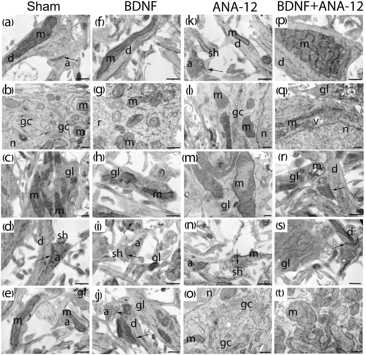
FIGURE 8.
Representative electron microscopy images of dissociated hippocampal cells on DIV 14. (a–e) Sham, (f–j) BDNF, (k–o) ANA-12, (p–t) BDNF + ANA-12. (a) Axo-spiny synapse; the cristae of mitochondria in a dendrite are smooth. (b) Cytoplasm of a neuron; mitochondria, Golgi apparatus, granular endoplasmic reticulum, numerous ribosomes and part of the nucleus are well visualized. (c) Mitochondria in a glial cell; cristae are smooth, and intermitochondrial contacts are visible. (d) Axo-dendritic and axo-spiny asymmetric synapse. (e) Immature axo-spiny synapse; no postsynaptic density is observed. (f) Mitochondrion in the neuronal dendrite. (g) Mitochondria in a neuronal soma; some are without cristae and have an enlightened matrix and short cristae. (h) Glial outgrowth with few glycogen pellets and irregular cristae. (i) Axo-spiny synapse; osmiophilic synaptic bubbles in an axon. (j) Axo-dendritic and axo-spiny asymmetric synapses, osmiophilic mitochondrion in an axon; mitochondria in the axonal outgrowth are destroyed. (k) Axo-spiny asymmetric synapse, short postsynaptic density. (l) Part of a neuronal soma, destroyed Golgi apparatus, few ribosomes on the endoplasmic reticulum, and normal mitochondrial structure. (m) Mitochondria with disrupted cristae in a glial cell. (n) Axo-spiny asymmetric synapse, osmiophilic bubbles among synaptic vesicles, mitochondrion with an irregular shape and high osmiophility. (o) Part of a neuronal soma, Golgi apparatus, and mitochondria with impaired structures. (p) Mitochondrial cluster in a dendrite. (q) Golgi apparatus with an increased area, a vacuole and destroyed mitochondria in the cytoplasm. (r) Axo-dendritic synapse and vacuolated mitochondria in a glial outgrowth. (s) Glial cell with a smooth endoplasmic reticulum, synaptic vesicles of different sizes in the axon, and mitochondrion with a reduced area. (t) Mitochondrial clusters in the neuronal cytoplasm; many of these clusters have extended cristae; mitochondria without cristae were also visualized. Scale bar – 0.5 μm.

