
Figure 3.
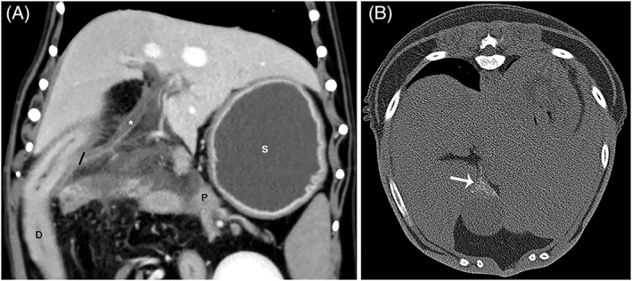
(A), Dorsal plane reconstruction in venous phase of biliary duct dilatation (*). The black line indicates the level of the insertion of the common bile duct on the duodenum. (B) Transverse non‐contrast image in a bone window with the dog in dorsal recumbency highlighting mineralization within the gall bladder (arrow). In image A, the pancreas (P) is seen immediately adjacent to the insertion of the common bile duct on the duodenum (D). Also notice the fluid filled stomach (S) indicating gastric ileus. Biliary mineralization was significantly more likely to be identified by CTA than ultrasound.