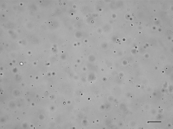
Figure 4.

SediVue image from a lowly cellular, unstained urine sediment from a dog containing many lipid droplets, where the image is focused on the lipid plane rather than on the few cells present. On manual microscopy, red and white blood cells were <1/high power field, and no epithelial cells or crystals were observed (scale bar = 50 μM)
