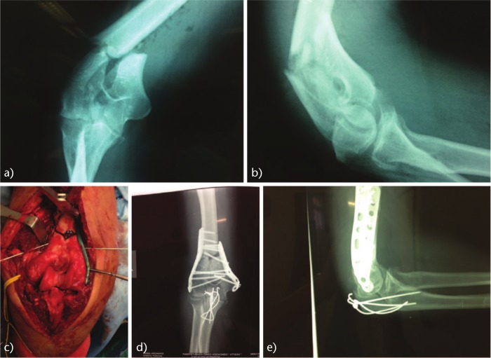
Fig. 1.
Fracture of the distal humerus in 52-year-old female treated with ORIF (double plating) with no proper surgical technique. A: anteroposterior radiograph of the distal humerus showing supra and inter-condylar fracture with comminution and B: lateral radiograph of the distal humerus showing supra and inter-condylar fracture with comminution. C: intraoperative image of the distal humerus fracture showing a gap in the metaphyseal area. D: anteroposterior radiograph of the elbow and E: lateral radiograph of the elbow seven months postoperative showing nonunion in the supracondylar area. The ulnar-medial plate is too short. The olecranon osteotomy is stabilized with tension-band technique; however, it is insufficiently fixed and not compressed.

