Fig. 2.
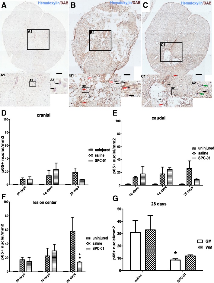
Immunohistochemical staining with hematoxylin and DAB (NF-kB p65) of the spinal cord from uninjured rats (a; A1, A2), 28 days after injury from rats treated with saline (b; B1, B2), or 28 days from rats transplanted with SPC-01 (c; C1, C2). Black arrows point to the nuclei void of NF-kB p65 expression, red arrows highlight the nuclei with translocated p65, and green arrows indicate cells with nuclei negative to p65 surrounded by p65+ cytoplasm (A1, A2, B1, B2, C1, C2). NF-κB nuclear translocation in the cranial and caudal areas did not differ significantly between the treatment groups (d, e). In the lesion center of the spinal cords, a significantly lower NF-κB p65 activity was observed at 28 days in the group treated with SPC-01 cells (f). A comparison of nuclear expression of NF-κB in the entire studied areas (gray and white matter) of injured spinal cords at day 28 after SCI showed lower p65 nuclear translocation in SPC-01-treated rats, which was more prominent in the gray matter of the injured spinal cords (g). Data are shown as the mean and SEM, *p ≤ 0.05, ** p ≤ 0.01. Scale bars, 200 μm

