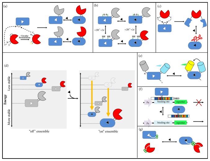
Figure 4.
Domain insertion strategies for converting PBPs into switchable proteins. (a) In a typical domain insertion, the inserted domain (red) has proximal N- and C-termini. These natural termini can be closed by a linker and new terminals can be created by circular permutation. The new termini of the circular permuted protein are inserted into a surface loop of the acceptor protein, PBP (blue). Combination of circular permutation and domain insertion increases the overall diversity of the protein switch library, generating different geometries. (b) Multi-input protein switches can be created by introducing disulfide bonds (green) to keep the PBP domain in an unbound (closed) conformation, which keeps the output domain in an “off” state. This example shows an AND gate logic in which the presence of both inputs, reduction of the disulfide bonds and ligand, is necessary to activate the output domain. (c) Mutually exclusive folding. The terminals of the inserted domain are far away from each other. This configuration creates a structural tug-of-war between the domains. Ligand binding stabilizes the PBP fold, which mechanically unfolds the output domain. (d) Ensemble model of allostery. In an “off” ensemble, the most probable chimera state has inactive input and output domains. Ligand binding remodels the population in the ensemble by increasing the stability of those states that bind the ligand and the most probable state has active domains. (e) FRET-based biosensors. FRET depends on the physical distance between a donor and an acceptor fluorophore. The PBP conformational change in response to ligand approximates the fluorescent proteins allowing energy transfer. (f) Inducible transcription factors can be designed through fusion between PBPs and DNA-binding domains (DBDs), leading to versions of DBDs that respond to new ligands. (g) Rational domain insertion can be possible through computational analysis (statistical coupling analysis-SCA). The analysis of the network of coevolving residues can predict distant sites on the surface. These sites can be used for coupling fusions. PBPs are shown as a rectangular (closed form, unbound) or an oval (open form, bound) shape. A grey color of the domain indicates that the protein is inactive. The signal that modulates the switch is showed as a black triangle.

