Fig. 1.
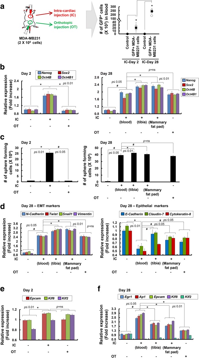
Analysis of tumor formation, transcriptional changes, and sphere-forming ability of MDA-MB231 cells harvested from the blood after intra-cardiac injection or from mammary fat pads after orthotopic injection. a Green fluorescent protein (GFP)+ MDA-MB231 cells (density, 2 × 10⁵ cells) were injected into the left ventricle of the heart or mammary fat pads of mice (n = 5). Right panel, the total bio-fluorescent GFP+ cells in the whole blood from the intra-cardiac (IC)-injected mice or PBS-injected control mice were isolated by fluorescence-activated cell sorting (FACS) and the number of GFP+ cells is presented. An average is shown as black circles; *p < 0.05. b Expression of stemness marker genes (Nanog, Sox2, Oct4B, and Oct4B1) was analyzed by quantitative real-time RT-PCR at 2 or 28 days following IC or orthotopic (OT) administration of MDA-MB231 cells (n = 5). In mice where MDA-MB231 cells were injected systemically (IC), secondary tumors formed in the tibia and mammary fat pads, and their expression of stemness marker genes was significantly increased (Tibia, Mammary fat pads). c Sphere-forming capacity of GFP+ MDA-MB231 cells harvested at 2 or 28 days from mice directly injected into left ventricle of the heart or orthotopically implanted into mammary fat pads (n = 5). d Expression of the epithelial-mesenchymal transition (EMT) marker (N-Cadherin, Twist, Snail1, and Vimentin; left panel) and epithelial marker (E-Cadherin, Claudin-7, and Cytokeratin-8) genes on GFP+ MDA-MB231 cells harvested at 28 days from blood, tibia, and mammary fat pads analyzed by quantitative real-time RT-PCR (n = 5). Expression of the shear stress (SS)-induced genes (Egr1, Ap1, Epcam, Klf8, and Klf2) on GFP+ MDA-MB231 cells harvested at 2 days (e) and 28 days (f) from blood, tibia, and mammary fat pads analyzed by quantitative real-time RT-PCR analysis (n = 5). Expression of each gene in quantitative real-time RT-PCR analysis was normalized to Gapdh. The data presented here are presented as mean ± SEM and are representative of three independent experiments. Statistically significant differences are tested at p < 0.05 significance

