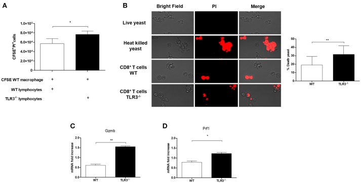
Figure 5.
Cytotoxicity of CD8+ T cells from TLR3−/− mice. (A) Cytotoxicity assay was performed. Macrophages derived by bone marrow of WT mice were stained with CFSE and infected with 1 × 10⁵ Pb18 yeast. After 4 h the lymphocytes WT or TLR3−/− were added in the culture (5 × 10⁵cells) and the cytotoxicity was measured with the dye PI by flow cytometry. Results are representative of three independent experiments. (*P < 0.05). Yeast activity of culture supernatants from CD8+ T cells exposed as in (B). Live yeast were cultured overnight with the supernatant and then incubated with dye PI before examination by fluorescence microscopy. (C) Relative expression of GZMB and (D) Prf1 by RT-PCR in CD8+T cells exposed to macrophages in (A). (*P < 0.05 and **P < 0.001).

