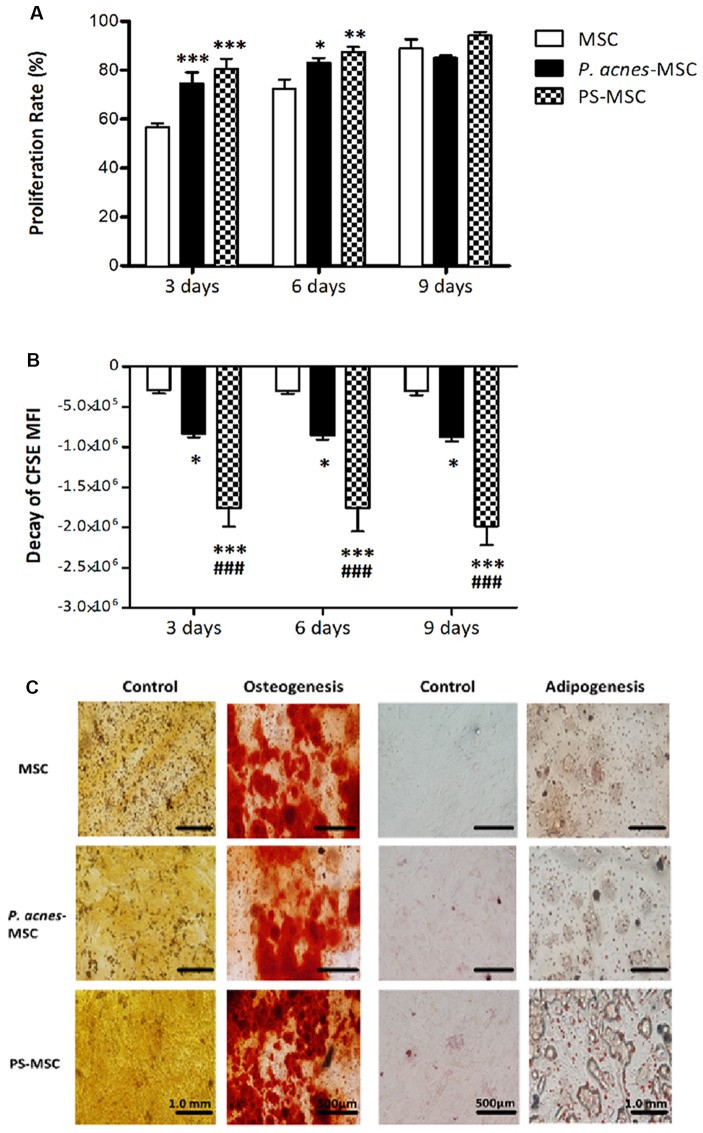
Figure 2.
P. acnes and PS treatment increase MSC proliferation without affecting differentiation. Cells obtained from P. acnes-, PS- and saline-treated groups were stained with CFSE and cultured for 9 days. (A) MSC proliferation rate analysis at 3, 6 and 9 days. (B) The decay of mean fluorescence intensity (MFI) proliferative cycles after 3, 6 and 9 days of culture. (C) MSCs were cultured under control, osteogenic or adipogenic conditions for 21 days. Osteogenic differentiation was observed after Alizarin Red S staining, and calcified nodules can be seen in red in all groups. Adipogenic differentiation was observed after Oil Red O staining, and lipid vesicles appear in red. Data are presented as the mean ± SEM of three mice per group, from two independent experiments. *p < 0.05, **p < 0.001 and ***p < 0.0001, when significance was calculated in relation to the MSC group, and ###p < 0.0001, when significance was calculated in relation to the P. acnes-MSC group, determined by two-way ANOVA followed by Bonferroni’s post-test.

