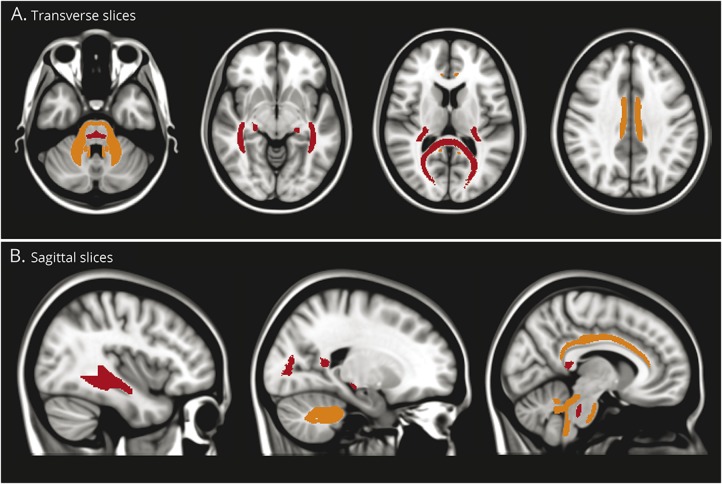
Figure 4. White matter tracts affected in essential tremor (ET) relative to Parkinson disease (PD).
Graphical representation of the location of the white matter tracts from the region of interest analysis where fractional anisotropy (FA) is lower (red) or radial diffusivity (RD) is higher (orange) in ET compared to PD. Lower FA could indicate demyelination or axonal degeneration while higher RD is more specific to demyelination. The white matter tracts with prominent differences appear to localize to cerebello-thalamo-cortical trajectories associated with the visual pathway.

