
Figure 1.
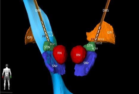
Bilateral DBS implantation sites. Target regions were STN and Field of Forel (FF) with pallido‐fugal fibers (not shown). Left corticospinal tract shown as light blue fibers. FF stimulation; left: case positive, EC4 negative, EC5 negative (1.4 mA, 0.6 mA; 90 us, 119 Hz), right: case positive, EC12 negative, EC13 negative (1.6 mA, 0.4 mA, 90 us, 119 Hz). STN stimulation was performed as follows; left: case positive, E2 negative, 1.8 mA, 90us, 119Hz, right case positive, EC10 negative, 1.8 mA, 90 us, 119 Hz (reduced frequency [60 Hz] did not lead to a better outcome, increased frequency and amplitude of STN DBS led to increased postural instability during gait).
Abbreviations: DBS, deep brain stimulation electrode; EC, effective contact; GPi, globus pallidus internus, (3D reconstruction with BrainLab Elements); RN, red nucleus; SNr, substantia nigra; STN, subthalamic nucleus.