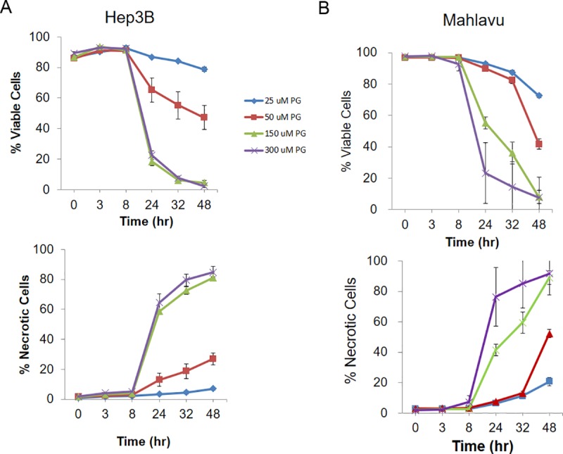
Fig 3. PG induces cell apoptosis in HCC cell lines.

The percentage of viable and necrotic cells was measured by high-content analysis at different time intervals. (A) The percentage of viable cells was decreased after PG treatment in Hep3B cells. The necrotic signals were increased after PG treatment in a dose-dependent manner. (B) In Mahlavu cells, the number of viable cells was significantly decreased, whereas necrotic cells were increased after PG exposure. The data are presented as the mean±SD of three independent experiments in triplicate.
