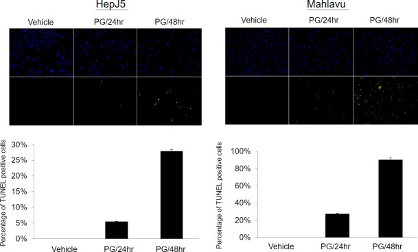
Fig 5. PG treatment induced cell apoptosis as monitored by a TUNEL assay.
(A) Representative fluorescent figure of the apoptotic assay. Blue DAPI was used to stain nuclei and TdT tagged with a green fluorochrome was used to detect apoptotic DNA fragmentation. There were few cells with positive apoptotic signals in the vehicle control sample. However, exposure to 80 μg/ml PG for 24hr or 48hr induced dramatically more apoptotic signaling in a time-dependent manner.

