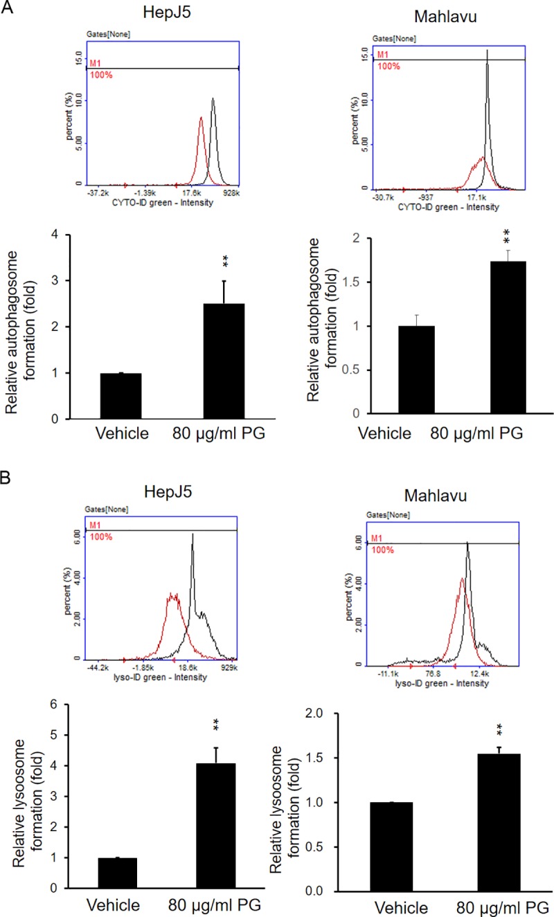
Fig 7. PG increases the formation of autophagosomes and lysosomes.

HepJ5 cells were exposed to 80 μg/ml PG for specific times. Autophagosome and lysosomes were detected using specific dyes and detected by NC-3000. (A) The formation of autophagosomes was increased after PG treatment in HepJ5 and Mahlavu cells compared with the vehicle treatment. (B) The formation of autophagosomes and lysosomes was increased after PG treatment compared with the vehicle treatment in HepJ5 and Mahlavu cells. The data are presented as the mean±SD of three independent experiments in triplicate (**p<0.01).
