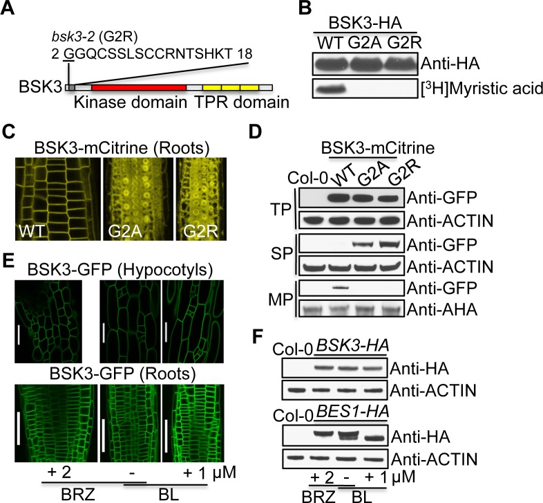
Fig 2. BSK3 is an N-myristoylated protein that localizes to the plasma membrane.
(A) A putative BSK3 N-myristoylation sequence GGQCSSLSCCRNTSHKT predicted by NMT, the MYR predictor. (B) In vitro myristoylation assays. (C) Subcellular localization of BSK3WT/G2A/G2R-mCitrine proteins. Root tip regions of 4-day-old light-grown seedlings were analyzed. (D) Western blots detecting BSK3WT/G2A/G2R-mCitrine proteins. Proteins were extracted from 7-day-old light-grown seedlings. Twenty to thirty micrograms of total proteins (TP), soluable proteins (SP), and microsomal proteins (MP) were loaded. AHA (Arabidopsis H+-ATPases) proteins were detected by an anti-AHA antibody. (E) Subcellular localization of BSK3-GFP protein. Seedlings were grown on 2 μM BRZ in the light for 4 days, or 4-day-old light-grown seedlings were treated with 1 μM BL for 2 hours. Scale bars = 50 μm. (F) Western blots detecting BSK3-HA and BES1-HA proteins. Seedlings were grown on 2 μM BRZ for 6 days, or 6-day-old light-grown seedlings were treated with 1 μM BL for 2 hours. Thirty micrograms of total proteins were loaded.

