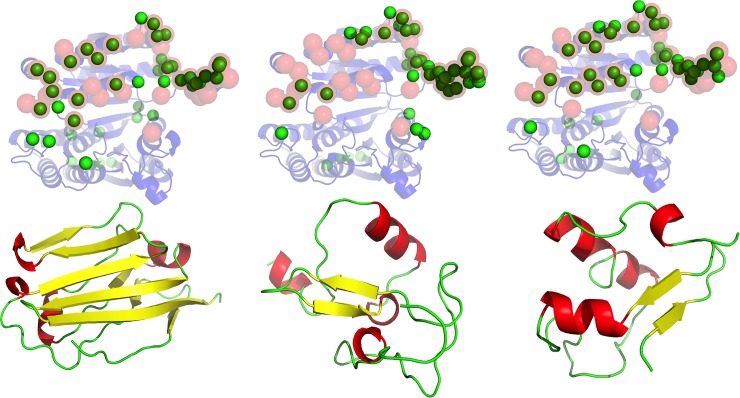
Fig 1. Binding supersite of 1cnz.A.
Three non-cognate ligands (lower row, from left to right, PDB codes: 2jjs.C, 2v86.A, 3h33.A) that share no detectable sequence or structure similarity to the cognate ligand, are docked extensively on the surface of the receptor (upper row, 1cnz.A). In the upper row, ribbon model in transparent blue shows the receptor structure. The annotated functional site in the receptor is shown using red transparent spheres for the Cα atoms. The predicted functional site residues, as defined by the corresponding ligand probes underneath, is shown using green spheres for the Cα atoms.

