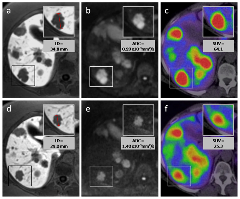
Fig. 3.
Responding lesion—a 54-year-old female with hepatic metastases from a pancreatic NET. After two cycles of intraarterial PRRT with 4 GBq 90Y- and 177Lu-DOTATOC, the longest diameter of the target lesion (box) decreased as can be seen on MR images with the hepatocyte-specific contrast agent Gd-EOB-DTPA (a, d). The volume of the lesion decreased from 11.0 to 5.0 cm3. On diffusion-weighted MRI (b, e) an increase of ADCmean from 0.99×10 to 1.40×10⁻³ mm2/s was measured, while the SUVmax on 68Ga-DOTATOC-PET/CT decreased from 64.0 to 25.3. Upper row of figures images before PRRT, lower row images after PRRT.

