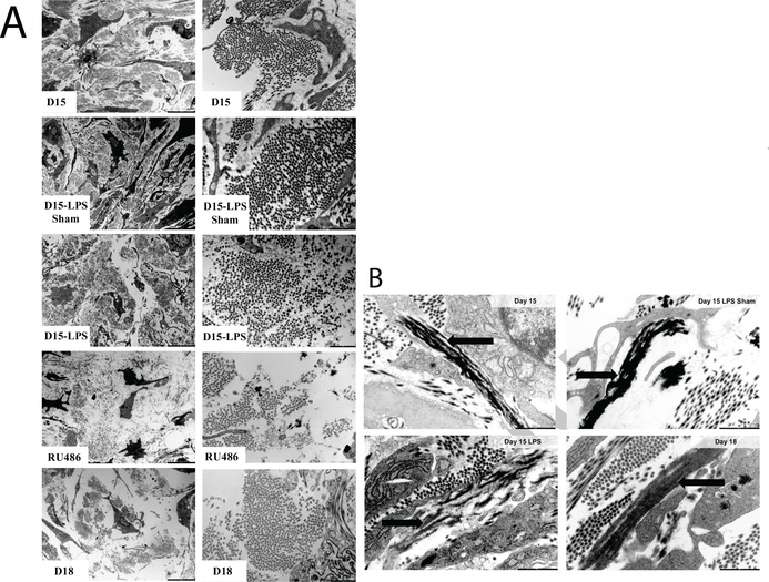
Figure 11: Ultrastructural assessment of cervical collagen and elastic fibers organization through transmission electron microscopy imaging.
a) Collagen fibers as seen on TEM images of cervical ECM taken on gestation d15, d15-LPS sham, d15-LPS, d15-RU486 (MFP) and d18. Left panel: 4200×; Right panel: 20500× magnification. Images reproduced from [15] with permission. b) Elastic fibers of cervical ECM observed through TEM for d15, d15-LPS sham, d15-LPS and d18 cervices. Black arrows indicate the elastic fibers ultrastructure which appears disrupted in d15-LPS, with elastin not being properly integrated in the microfibrillar scaffold of the elastic fibers compared to control. Scale bars are 1m. Images reproduced from [24] with permission.

