. 2019 Jan 7;8:e41741. doi: 10.7554/eLife.41741

Figure 5. Conformation of the Schiff base region of GtACR1.

(A–B) Structural comparison shows different H-bond networks (dashed lines) in GtACR1 (A) and C1C2 (B). (C) the H-bond network in the ENS triad of GtACR1. The tunnel (black dots) assessed by CAVER. (D) Peak photocurrents generated by the wild-type GtACR1 and indicated mutants in response to laser flash excitation. The black squares, mean; line, median; box, SE; whiskers, SD; empty diamonds, raw data recorded from individual cells.

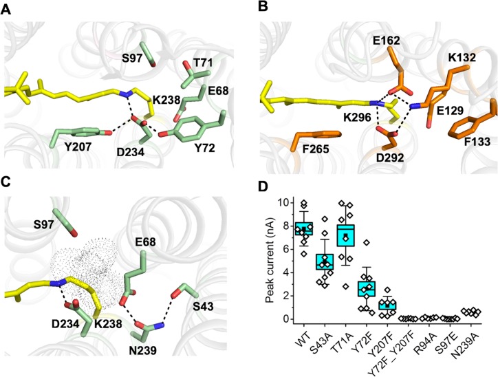
Figure 5—source data 1. Numerical data for the current amplitude values measured in individual cells are shown in Figure 5D.
DOI: 10.7554/eLife.41741.034

Figure 5.

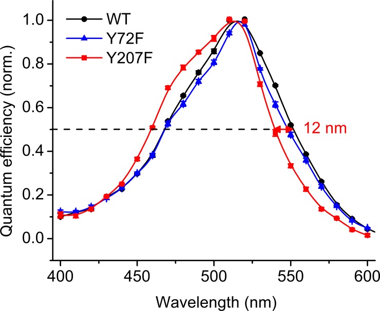
Figure 5—figure supplement 1. The action spectra of photocurrents generated by the Y72F and Y207F mutants, as compared to the wild type.

Figure 5—figure supplement 1.

The data points are mean values of 4–6 individual scans. For more detail see Materials and methods.
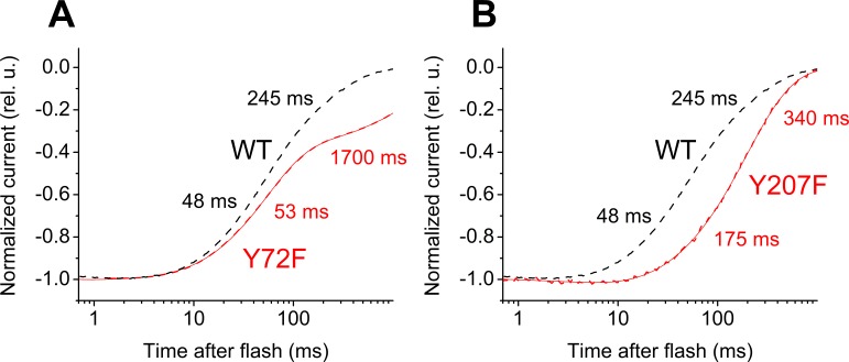
Figure 5—figure supplement 2. Laser flash-evoked photocurrents generated by the Y72F and Y207F mutants, as compared to the wild type.

Figure 5—figure supplement 2.

Thin solid lines are recorded traces, and thick dashed lines are multiexponential fits. The numbers are the time constants of individual decay phases.
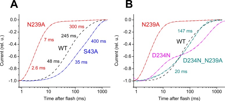
Figure 5—figure supplement 3. Laser flash-evoked photocurrents generated by.

Figure 5—figure supplement 3.

(A) the S43A and N239A mutants, as compared to the wild type; (B) the D234N, N239A and D234N_N239A_ mutants, as compared to the wild type. Thin solid lines are recorded traces, and thick dashed lines are multiexponential fits. The numbers are the time constants of individual decay phases.
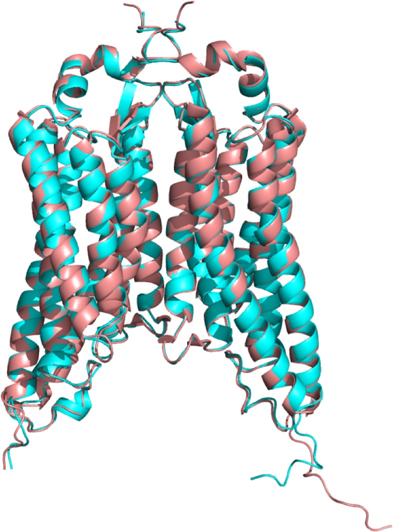
Figure 5—figure supplement 4. Superposition of two independently obtained GtACR1 structures.

Figure 5—figure supplement 4.

Overview of the GtACR1 structure (PDB code: 6EDQ) colored in cyan superimposed on the structure of GtACR1 reported by Kim et al., 2018 (PDB code: 6CSM) in light pink.
The RMSD value between these two structures is ~0.4 Å.