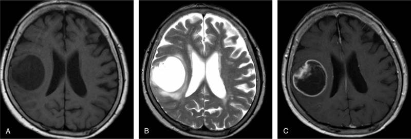
Figure 1.

Neuroradiological features. A frontoparietal tumor exhibiting a neuroradiological cyst with mural nodule pattern on magnetic resonance imaging. The mural nodule is hypointense in the T1-weighted (A) and hyperintense in the T2-weighted (B) images. T1-weighted gadolinium-enhanced image reveals the brain tumor as a heterogeneously enhancing mural nodule with a rim-enhancing cyst (C).
