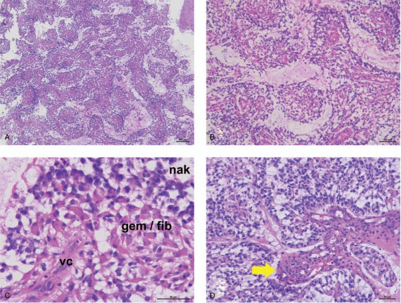
Figure 2.

Histopathological features. Histopathologically, the highly cellular tumor exhibits pseudopapillary structures with non-hyalinized central capillaries (A and B; hematoxylin and eosin [H&E], magnification ×40 [A] and ×100 [B]). Non-hyalinized vascular cores (vc) surrounded by both fibrillary (fib) and mini-gemistocytic (gem) neoplastic cells; these pseudopapillary structures are covered by naked neoplastic cells (nkd) (C; H&E, magnification ×400). Focal microvascular proliferation is also observed (D; H&E, magnification ×200).
