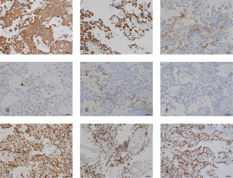
Figure 3.

Immunohistochemical features. Immunohistochemically, neoplastic cells express glial fibrillary acidic protein (A; magnification ×200), and oligodendrocyte transcription factor 2 (B; magnification ×200). Tumor stroma reveals immunoreactivity against synaptophysin (C; magnification ×200). A few residual neurons in the tumor show neuronal nuclei (NeuN) immunopositivity (D; ×200), and neurofilament immunoreactivity is focally observed in the tumor stroma (E; magnification ×200). The tumor exhibits no immunoreactivity against mutant isocitrate dehydrogenase (IDH1) R132H (F; magnification ×200) and retains nuclear expression of α-thalassemia/mental retardation syndrome X-linked (G; magnification ×200) and INI-1 (H; magnification ×200). Additionally, MIB-1 labeling index is very high (63.4%) (I; magnification ×100).
