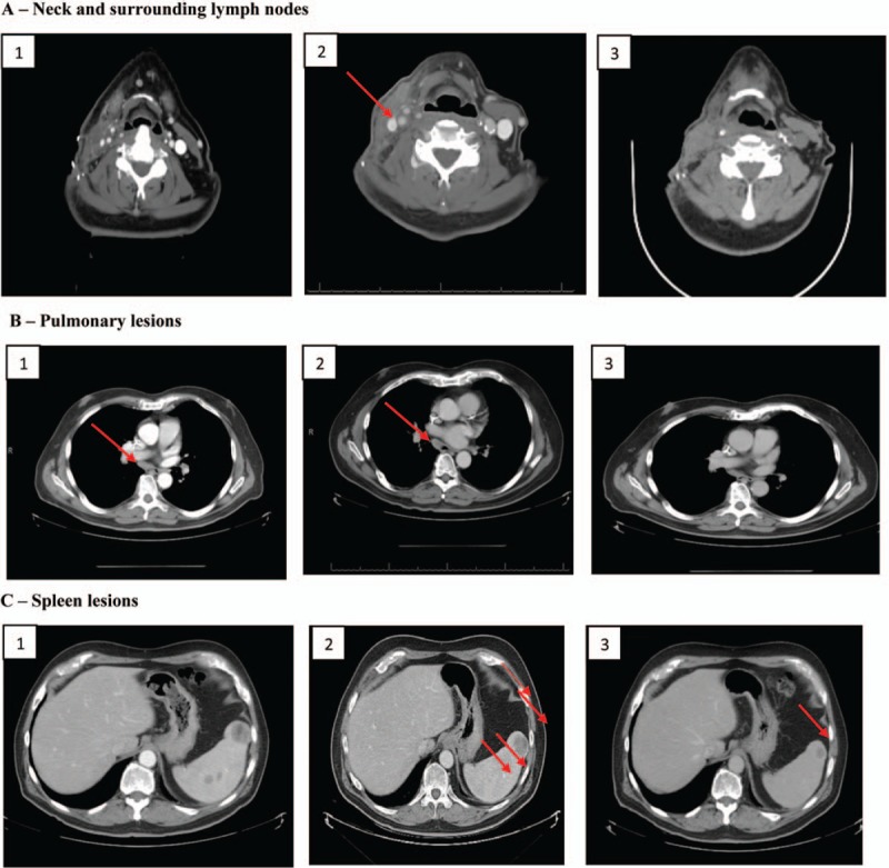
Figure 2.

Response to therapy monitored by computed tomography. (A) Neck lesion and surrounding lymph nodes: (1) Before BRAFi treatment (January 2014). (2) Recurrence of melanoma after BRAFi treatment (December 2014). (3) After anti-CTLA-4 treatment (April 2015). (B) Pulmonary lesions: (1) After nivolumab and before pembrolizumab treatment (August 2015). (2) During pembrolizumab treatment (March 2016). (3) During pembrolizumab treatment (April 2018). (C) Spleen lesions: (1) Before pembrolizumab treatment (October 2015, size 3.5 cm). (2) During pembrolizumab treatment. (March 2016, size 3 cm). (3) During pembrolizumab treatment (April 2018, size 1.5 cm).
